
After the immobilization period has ended, the cast will be removed and further analysis of the injury will be required. The duration of the immobilization is at the treating physician's discretion. A long arm cast may be required in order to ensure that all wrist movement has been stopped. Conservative treatments include immobilization and stabilization of the affected wrist by placing it in a cast. After the ECU injury is diagnosed, a physician will choose a course of treatment, which depends upon the severity of the injury. Īn ECU injury most often requires imaging (CT, MRI, ultrasound) for diagnosis. A similar injury involving the medial elbow is known as golfer's elbow.

Some treatments for tennis elbow include occupational therapy, physical therapy, anti-inflammatory medication, and rest from the activity that caused the injury. As a result, this causes irritation to the already existing injury. This pain intensifies because the extensor carpi ulnaris has an injury near the elbow area and as a person moves their arm, the muscle contracts, thus causing it to move over the medial epicondyle of the humerus. The pain worsens when a person moves their wrist with force. Some symptoms of an extensor carpi ulnaris injury include pain when shaking hands or when squeezing/gripping an object. This injury occurs in people who participate in activities requiring repetitive arm, elbow, and wrist, especially when they are tightly gripping an object. Injuries Ī common injury to the extensor carpi ulnaris is tennis elbow. It would therefore be paralyzed in an injury to the posterior cord of the brachial plexus. Innervation ĭespite its name, the extensor carpi ulnaris is innervated by the posterior interosseous nerve (C7 and C8), the continuation of the deep branch of the radial nerve. In this case it would be described as ulnaris lateralis. The muscle is a minor extensor of the carpus in carnivores, but has become a flexor in ungulates. The extensor carpi ulnaris extends the wrist, but when acting alone inclines the hand toward the ulnar side by its continued action it extends the elbow-joint. It originates from the lateral epicondyle of the humerus and the posterior border of the ulna, and crosses the forearm to the ulnar (medial) side to insert at the base of the 5th metacarpal. The extensor carpi ulnaris acts to extend and adduct at the carpus/wrist from anatomical position.īeing an extensor muscle, extensor carpi ulnaris is located on the posterior side of the forearm. The operation involves releasing the extensor retinaculum that forms the roof of the first dorsal compartment and any surrounding septa.In human anatomy, the extensor carpi ulnaris is a skeletal muscle located on the ulnar side of the forearm. If conservative measures fail to relieve the symptoms, then operative management can be pursued. Nonoperative treatment is successful in up to 80% of patients.

First-line treatment is nonoperative management with rest, thumb spica splinting, NSAIDs, and corticosteroid injections. Radiographs can help eliminate other causes of radial-sided wrist pain however, the diagnosis of De Quervain’s syndrome is clinical. Furthermore, provocative tests reproduce the patient’s symptoms. On physical examination, patients have tenderness to palpation over the APL and EPB tendons at the radial styloid. Patients complain of wrist pain that worsens with grasping or twisting motions. The syndrome is common in middle-age women and overuse injuries.

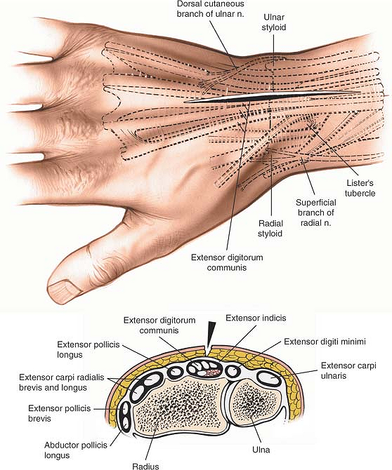
De Quervain’s syndrome is a frequent cause of radial-sided wrist pain that emanates from a narrowing of the first dorsal compartment due to synovial inflammation. The extensor tendons of the wrist are divided into six compartments based on synovial sheaths that extend from the overlying extensor retinaculum.


 0 kommentar(er)
